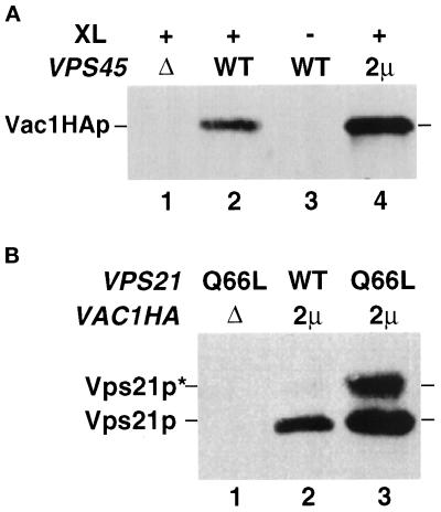
Figure 3.
Vac1p interacts with Vps45p and Vps21p in vivo. (A) Spheroplasts (5 OD600 equivalents) of the following strains were prepared: GTY107 (Δvps45Δvac1) expressing Vac1HAp (lane 1), GTY104 (Δvac1) expressing Vac1HAp (lanes 2 and 3), and GTY104 expressing Vac1HAp and overexpressing Vps45p (lane 4). The spheroplasts were lysed, and the lysates were treated with (+) or without (−) the reducible cross-linker DSP (XL) as described. Vps45p and cross-linked proteins were immunoprecipitated with Vps45p antibodies. The immunoprecipitates were resolved by SDS-PAGE and transferred to nitrocellulose, and the blots were probed with HA antiserum. Vac1HAp was visualized using Blaze chemilluminescent detection reagents. (B) Spheroplasts of GTY112 (Δvac1Δvps21) (25 OD600 equivalents) overexpressing Q66L-Vps21p (lane 1), Vps21p and Vac1HAp (lane 2), or Q66L-Vps21p and Vac1HAp (lane 3) were lysed and centrifuged at 16,000 × g. HA antisera (10 μl) was added to the cleared supernatants and incubated for 30 min at 4°C. Protein G-Sepharose was added, and the reactions were incubated an additional 30 min at 4°C. Sepharose–antibody–antigen complexes were pelleted at 2000 × g and washed five times with native lysis buffer. Proteins were eluted with sample buffer, resolved by SDS-PAGE, and subjected to Western analysis using Vps21p antisera. Prenylated (22 kDa, Vps21p) and unprenylated (23 kDa, Vps21p*) Vps21p were detected using Blaze chemilluminescent detection reagents.

