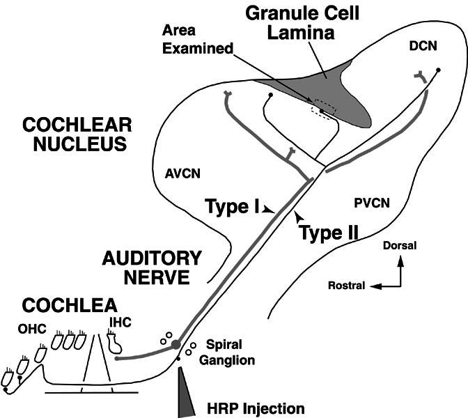
Figure 1.

Schematic showing the site of horseradish peroxidase (HRP) injection to label type II fibers in the spiral ganglion of the cochlea. Auditory nerve fibers are shown peripherally in the cochlea where type II fibers form the afferent innervation of the outer hair cells (OHC) and type I fibers form the afferent innervation of the inner hair cells (IHC). Both fiber types project centrally in the auditory nerve, bifurcate in the cochlear nucleus, and form branches in the anteroventral, posteroventral, and dorsal subdivisions of the cochlear nucleus (AVCN, PVCN, and DCN). Dividing the VCN from the DCN is the granule-cell lamina, a region of termination for many type II fibers. Type II fibers in the lamina were examined in the approximate location indicated by the dashed box. The figure orientation is approximately the sagittal plane (see compass indicating dorsal toward the top of the figure). In successive figures, the orientation is reversed (dorsal toward the bottom of the figure) in order to be consistent with our previous work (Brown et al. 1988b; Benson and Brown 1990; Benson et al. 1996).
