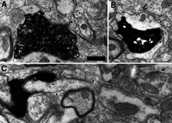
Figure 3.

Electron micrographs of three small dendrites (delineated with small arrowheads) receiving synapses from type II fibers. In panels A and C the postsynaptic density is indicated with large arrowheads, but in panel B the labeled terminal was synaptic in other sections. In panel A, the small dendrite could not be traced to its soma of origin, but it received a second synapse from the same labeled terminal onto a spine in a nearby section (Fig. 2). The small dendrite of panel B became much larger in nearby sections, as if swollen relative to other parts of the dendrite. This dendrite was followed through serial sections to the soma of a small cell (Figs. 4, 5A). The small dendrite in panel C is sectioned relatively longitudinally and is seemingly engulfed by the labeled type II terminal, a swelling found at a branch point of the fiber. The dendrite in C was also connected, through serial sections, to a small cell (Fig. 5B). Arrow denotes density of a synapse formed by an unlabeled terminal containing small round vesicles. Scale bar in A = 0.5 μm and also applies to B and C.
