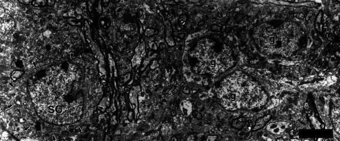
Figure 4.

Electron micrograph of a small cell (SC) and a nearby cluster of granule cells (g). The small cell received, on its small dendrite, a synapse from an en passant swelling of a type II axon (Figs. 3B, 5A). Like granule cells, the small cell has chromatin aggregates in its nucleus, but it is larger than the granule cells and has a distinctive nuclear fold. Its perikaryal organelles are more densely packed, thus its cytoplasm appears darker than that of the granule cells. Scale bar = 5 μm.
