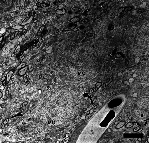
Figure 6.

Two presumed multipolar cells (MC) that received axosomatic synapses from labeled type II terminals. Swellings of the labeled type II fiber are indicated by two black, curved arrows. The swelling at the lower middle of the micrograph is a terminal swelling of the type II fiber and the swelling at the upper middle of the micrograph is a branch point swelling of the same type II fiber. This latter swelling synapses with a bootlike somatic appendage of the upper cell (outlined by small arrows). The cells are on the border of the lamina; a granule cell (g) is visible at lower right. Also at lower right, curving under the granule cell, is another labeled type II fiber (small arrows) illustrating the typical thin diameter of these fibers. Thicker-labeled myelinated fibers (m) and terminals from type I fibers within the core of the cochlear nucleus are visible at upper left. Scale bar = 5 μm.
