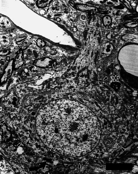
Figure 7.

Electron micrograph of a large neuron (outlined by arrows) that received two axosomatic synapses from a terminal swelling of a type II fiber. The neuron was located at the border of the granule-cell lamina, and it gave off a large dendrite directed toward the core of the ventral cochlear nucleus. This dendrite receives several terminals, one of which is darkened with HRP label. Another labeled axosomatic terminal is visible just beneath the asterisk. The origin of that terminal could not be determined unambiguously, but it connected with other axosomatic terminals that also formed punctate synapses with the neuron. Together, the terminals comprise a modified endbulb, suggesting that this neuron is a globular bushy cell that receives input from both type I and type II auditory nerve fibers. Scale bar = 5 μm.
