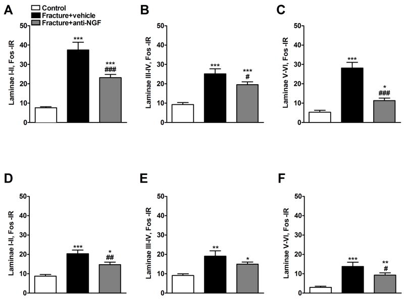
Figure 6.
Four weeks after tibia fracture and casting the L4, L5 lumbar spinal cord segments were sectioned and stained for Fos immunoreactivity. Results are expressed as the mean number of spinal Fos-IR neurons/section (± SEM), per dorsal horn laminar region (laminae I + II, III + IV and V + VI). Panels (A), (B), and (C) represent the right side of the spinal cord (ipsilateral to the fracture side), whereas panels (D), (E), and (F) represent the left side of the spinal cord (contralateral to the fracture side). There was a significant bilateral increase in Fos expression through dorsal horn laminae I-VI after fracture. Treatment with anti-NGF antibodies (n = 9, Fracture + anti-NGF) significantly decreased fracture induced spinal Fos expression, compared to the vehicle treated fracture rats (n = 6, Fracture+vehicle) through laminae I-VI ipsilaterally (A, B and C) and in laminae I-II and V-VI contralaterally (D and F). *P < 0.05, **P < 0.01, ***P < 0.001 fracture vs. control group (n = 6), and #P < 0.05, ##P < 0.01, ###P < 0.001 fracture anti-NGF antibodies vs. fracture vehicle treatment.

