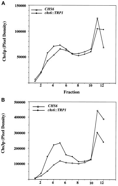
Figure 3.
Velocity gradient analysis of membranes from chs6 cells. Z130–1D (chs6) cells were converted to spheroplasts and lysed, and a 500 × g supernatant fraction was sedimented on a linear Ficoll gradient for 2 h. Gradient fractions were collected from the top and analyzed as in Figure 2. (A) Fractions were analyzed for Chs1p. (B) Fractions were analyzed for Chs3p.

