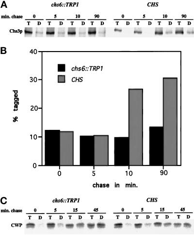
Figure 5.
CHS6-dependent and time-dependent cell surface labeling of newly synthesized Chs3p. (A) Strains Z130–1D (chs6) and Z130–1A (CHS) were labeled with 35S-Promix for 3 min at 25°C and chased for the indicated times. After cell surface proteins were tagged with TNBS, cells were lysed and immunoprecipitated with anti-Chs3p Abs, and the resulting precipitate was divided in half and either reprecipitated with anti-Chs3p Abs (T) or anti-DNP Abs (D) and analyzed by SDS-PAGE. (B) Quantification of (A) after PhosphorImager analysis. (C) Strains Z130–1D (chs6) and Z130–1A (CHS) were analyzed as above, but metabolic labeling was for 4 min and anti-CWP Abs were used instead of anti-Chs3p Abs.

