
Figure 4.
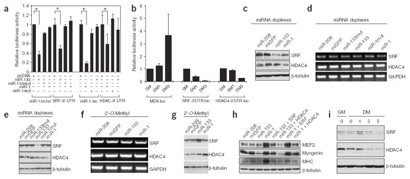
Identification of miR-1 and miR-133 target genes in skeletal muscle. (a) Repression of SRF and HDAC4 3′ UTRs by miR-133 and miR-1. Luciferase reporters containing either miR-133 complementary sites from mouse SRF 3′ UTR (SRF-3′-UTR), miR-1 complementary sites from mouse HDAC4 3′ UTR (HDAC4-3′-UTR) or the perfect antisense sequences of miR-133 (miR-133-luc) or miR-1 (miR-1-luc) were cotransfected with the indicated miRNA expression vectors. Luciferase activity was determined 48 h after transfection. Data represent the mean ± s.d. from at least three independent experiments done in duplicate (*P < 0.05). (b) SRF-3′-UTR, HDAC4-3′-UTR and MCK-luc luciferase reporters were transfected into C2C12 myoblasts. Cells were maintained in growth medium for 24 h (GM) or transferred into differentiation medium for 1 d (DM1) or 3 d (DM3) before luciferase activity was determined. (c–e) C2C12 myoblasts cultured in growth medium were electroporated with the indicated miRNA duplexes (or their mutants), or miR-208 and miGFP as controls. Cells were cultured in growth medium for 24 h after transfection before being either subjected to immunoblotting with anti-SRF and anti-HDAC4 antibodies (c), or transferred into differentiation medium for 24 h and subjected to RT-PCR for the indicated genes (d) or to immunoblotting with the indicated antibodies (e). (f,g) C2C12 myoblasts cultured in growth medium were electroporated with the indicated 2′-O-Methyl antisense oligonucleotide inhibitors. Cells were cultured in growth medium for 24 h after transfection and transferred into differentiation medium for 24 h before being subjected to RT-PCR for the indicated genes (f) or to immunoblotting with indicated antibodies (g). (h) C2C12 myoblasts cultured in growth medium were electroporated with the indicated miRNA duplexes and/or expression plasmids for SRF or HDAC4, as indicated. Cells were cultured in growth medium for 24 h after transfection. Immunoblotting with the indicated antibodies was done 24 h after transfer into differentiation medium. (i) C2C12 myoblasts were cultured in growth medium or differentiation medium for 0, 1, 3 or 5 d and subjected to immunoblotting with the indicated antibodies.