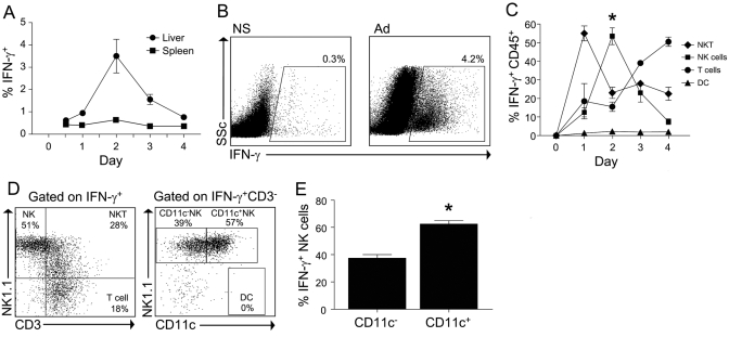
Fig. 1.
CD11c+ NK cells contribute to early hepatic IFN-γ production during adenovirus infection. Following infection with adenovirus, liver CD45+ cells and splenocytes were isolated at the indicated time-points and cultured for 6 h in the presence of Brefeldin-A without restimulation. (A) The percentage of liver leukocytes and splenocytes that were IFN-γ+ is shown. (B) Representative FACS plots of liver CD45+ cells isolated from animals treated 48 h previously with normal saline (NS) or adenovirus (Ad). SSc, Side-scatter. (C) The composition of IFN-γ+ cells at serial time intervals during adenovirus infection was determined by intracellular staining. The number of NKT cells (NK1.1+CD3+), NK cells (NK1.1+CD3−), DC (NK1.1−CD11c+CD3−), and conventional T cells (NK1.1−CD3+) is expressed as a percentage of total IFN-γ+ cells. (D) Representative FACS plots to demonstrate the gating strategy used to determine the composition of the various IFN-γ+ cell types. (E) The expression of CD11c on IFN-γ+ liver NK cells is shown at 48 h after adenovirus infection. Data represent the mean (±sem) of two separate experiments in which the hepatic leukocytes of three to four mice per group were pooled. *, P < 0.05.

