Fig. 2.
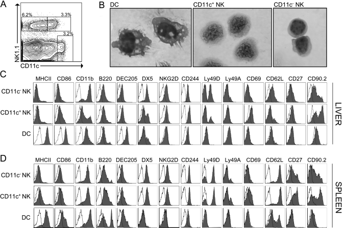
CD11c+ NK cells comprise a substantial portion of NK1.1+CD3− cells in the uninfected liver. (A) Liver NPC from unmanipulated mice were enriched with anti-CD45 immunomagnetic beads and analyzed by flow cytometry. The control-plot is gated on CD3− hepatic leukocytes to highlight conventional liver DC (NK1.1−CD11chiCD3−), CD11c+ NK cells (NK1.1+CD11c+CD3−), and CD11c− NK cells (NK1.1+CD11c−CD3−). The percentage indicates the fraction of CD3− cells. (B) FACS-purified DC, CD11c+ NK cells, and CD11c− NK cells were stained with H&E after cytospin and imaged at 100× original magnification. Similar results were obtained from two separate experiments, and the microscopic appearances of each cell population were greater than 90% consistent. (C and D) CD45+ liver cells and splenocytes from uninfected mice were analyzed for the indicated surface markers by flow cytometry after gating on DC, CD11c+ NK cells, and CD11c− NK cells. Closed histograms represent staining of the indicated surface markers, and open histograms represent isotype controls. Data are representative of a minimum of three separate experiments.

