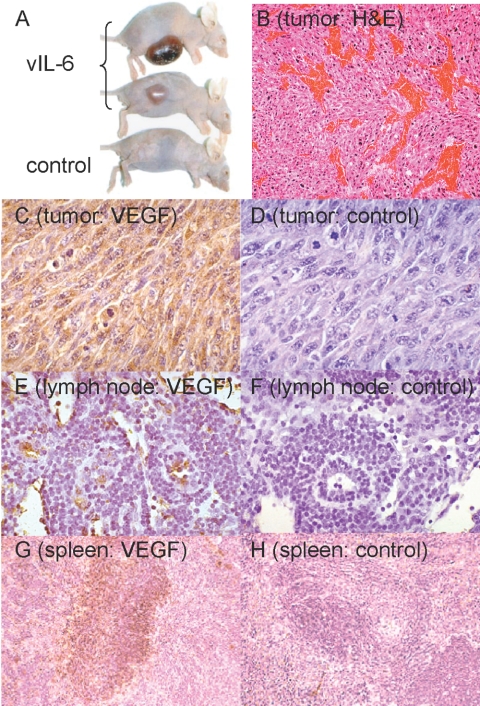
Fig. 2.
Tumorigenicity of NIH3T3 cells overexpressing vIL-6 in immunodeficient mice. (A) Representative tumors in nude mice 4 weeks after they were injected s.c. with vIL-6-NIH3T3 cells. Control NIH3T3 and vIL-6-NIH3T3 cells were injected at 0.5 × 10⁶ cells/mouse (BALBc nude). (B) Increased vascularization in vIL-6-NIH3T3 tumors. Representative image from tumor tissue stained with H&E (original magnification, ×100). (C, E, and G) VEGF visualized by immunohistochemical staining in tumor tissue (original, ×40), lymph node (original, ×200), and spleen (original, ×100), respectively. (D, F, and H) Control immunostaining of tumor tissue, lymph node, and spleen, respectively.

