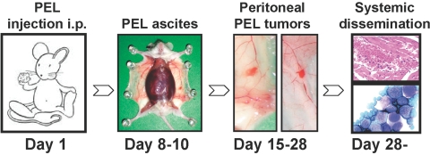
Fig. 6.
Schematic representation of a PEL mouse model. Immunodeficient NOD/SCID mice are injected i.p. with PEL cells (BC-1 cells, 20×106/mouse) on Day 1. On Days 8–10, the mice develop a lymphomatous ascite; in the example shown, the peritoneal cavity is closed. On Days 15–18, 50–75% of the mice develop visible tumors attached to the peritoneal mesothelium. From Day 29 on, most mice have evidence of PEL dissemination outside the peritoneal cavity. In the example shown, PEL cells are identified microscopically under the renal capsule and in the blood.

