Figure 6.
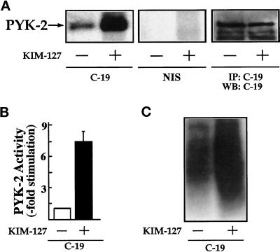
KIM-127 induces stimulation of PYK-2 tyrosine kinase activity. (A) T lymphoblasts plated on ICAM-1-Fc–coated plastic dishes were allowed to adhere for 30 min in the absence (−) or presence (+) of 10 μg/ml mAb KIM-127. The cells were then lysed, the extracts were incubated with C-19 antibody to immunoprecipitate PYK-2 (C-19) or with nonimmune goat serum as a control (NIS), and kinase reactions were performed as described in MATERIALS AND METHODS. PYK-2 levels were determined by immunoprecipitation (IP) with the C-19 anti-PYK-2 antibody and Western blot analysis (WB) with C-19 anti PYK-2 antibody. The position of PYK-2 is indicated with an arrow. The results shown are representative of seven independent experiments. (B) Quantification by densitometric scanning of the effect of KIM-127 on PYK-2 activity. PYK-2 was immunoprecipitated with the C-19 antibody and an in vitro kinase reaction carried out as described in MATERIALS AND METHODS. Values are the mean ± SEM of five independent experiments and are expressed as fold stimulation above control. (C) In vitro kinase reactions of the C-19 PYK-2 immunoprecipitates were performed in the presence of the tyrosine kinase substrate poly-Glu-Tyr (4:1). Phosphorylation of poly-Glu-Tyr (4:1) was carried out after a 15-min incubation with [γ-32P]ATP as described in MATERIALS AND METHODS. A representative experiment is shown.

