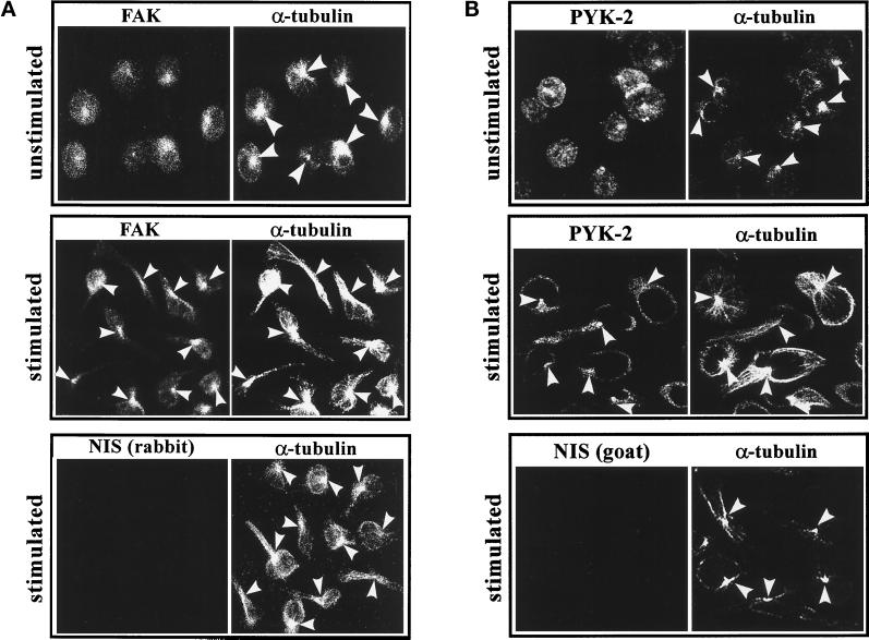
Figure 8.
FAK and PYK-2 colocalize with the MTOC in polarized lymphoblasts. Lymphoblasts were allowed to adhere onto ICAM-1-Fc–coated dishes for 60 min either in HEPES-NaCl buffer containing 1 mM Mg2+ and 1 mM Ca2+ (unstimulated) or in HEPES-NaCl containing 10 mM Mg2+ and 1 mM EGTA (stimulated). (A) The cells were fixed, permeabilized, and double stained with C-20 rabbit polyclonal antibody against FAK (FAK) or with a control rabbit nonimmune serum (NIS rabbit) and with an anti-α-tubulin mAb (α-tubulin) as specified in MATERIALS AND METHODS. (B) The cells were fixed, permeabilized, and doubled stained with C-19 goat polyclonal antibody against PYK-2 (C-19) or with a control goat nonimmune serum (NIS goat) and with an anti-α-tubulin mAb (α-tubulin) as specified in MATERIALS AND METHODS. Arrowheads indicate the positions of the MTOC in representative cells.

