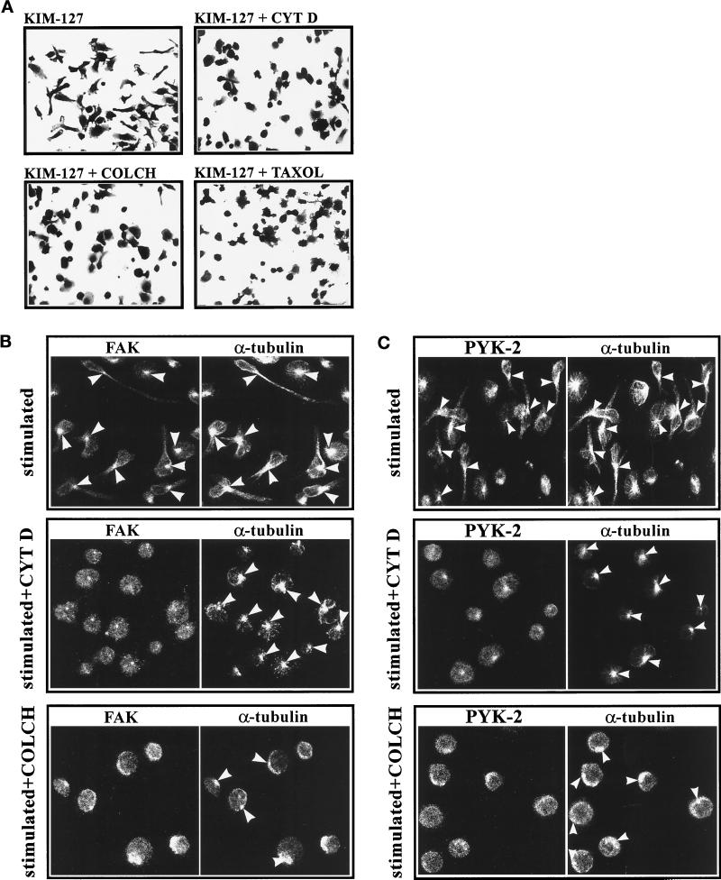
Figure 9.
Cytoskeletal interfering agents block LFA-1–dependent polarization of lymphoblasts and reduce translocation of FAK and PYK-2 to the MTOC. (A) T Lymphoblasts were washed in RPMI 1640 medium and then pretreated for 4 h with 0.3% DMSO (KIM-127), with 2.5 μM cytochalasin D dissolved in DMSO (KIM-127 + CYT D), with 10 μM colchicine dissolved in DMSO (KIM-127 + COLCH), or with 1 μM taxol dissolved in DMSO (KIM-127 + TAXOL). Cells were subsequently plated on ICAM-Fc–coated dishes and stimulated for 60 min with 10 μg/ml mAb KIM-127. Attached cells were fixed and stained with 0.5% crystal violet in 20% methanol before photography. A representative experiment is shown. (B and C) T Lymphoblasts were washed in RPMI medium and then pretreated for 4 h with 0.3% DMSO (stimulated), with 2.5 μM cytochalasin D (stimulated + CYT D), or with 10 μM colchicine (stimulated + COLCH). After this pretreatment, cells were washed in HEPES-NaCl buffer, plated on ICAM-1-Fc–coated dishes, and stimulated for 60 min with 10 mM Mg2+ plus 1 mM EGTA. The cells were fixed, permeabilized, and doubled stained with C-20 rabbit polyclonal antibody against FAK (B, FAK) or with C-19 goat polyclonal antibody against PYK-2 (C, PYK-2) and with an anti-α-tubulin mAb (B and C, α-tubulin). Immunofluorescence samples were analyzed by confocal laser microscopy as specified in MATERIALS AND METHODS. Arrowheads indicate the position of the MTOC. A representative experiment is shown.

