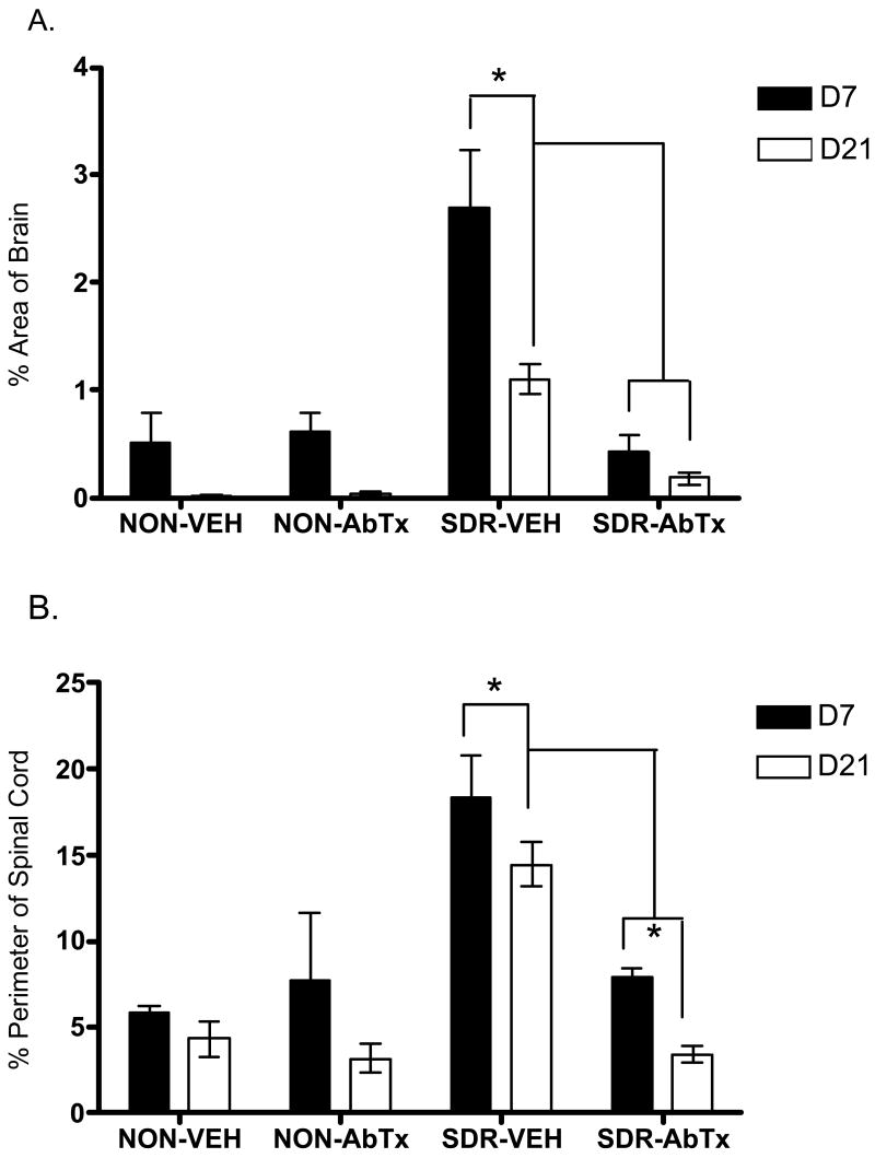
Fig 6.
Inflammation in brain (A) and in spinal cord (B) was elevated by SDR (SDR-Vehicle) across days 7 and 21 pi, but this SDR-induced increase in inflammation was prevented by IL-6 neutralizing antibody treatment (SDR-AbTx). Inflammation in brain is expressed as the percent of area with microgliosis and perivascular cuffing, whereas inflammation in spinal cord is expressed as the percent of section perimeter with meningitis. Significant post hoc differences are indicated by asterisks (*) for comparisons across groups and by “†” for comparisons of the neutralizing antibody condition collapsing across day pi.

