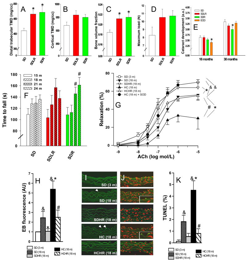
Figure 3. Resveratrol improves the health of mice fed a standard diet.
Femurs were removed following natural deaths between the ages of 30–33 months and analyzed by micro-computed tomography (micro CT). (A) Distal trabecular tissue mineral density (TMD) was improved in SDLR and SDR compared to SD control bones (P < 0.05 for SDLR and P < 0.01 SDR vs SD control; *). All error bars indicate s.e.m. (B) Cortical TMD tended to increase with resveratrol treatment (p = 0.14). All error bars indicate s.e.m. (C) Resveratrol significantly increased bone volume to total volume ratio over the entire femur in SD fed mice (P < 0.05 for SDLR and P < 0.01 SDR vs SD control; *), n = 5 for all groups in panels A, B, and C. All error bars indicate s.e.m. (D) Bone strength was tested in the post-mortem femurs. Resveratrol treatment caused a trend towards increased maximum load (the load that is endured by a bone prior to it failing in the three-point bending to failure test) (p = 0.18), n = 5 for SD and SDLR and n = 4 for SDR. All error bars indicate s.e.m. (E) Resveratrol treatment delayed the onset of age-related cataracts. Lens opacity was scored in living mice on a scale from 0 to 4 by half steps of 0.5, with 4 representing the complete lens opacity of a mature cataract. Age-related cataract development was significantly decreased at 30 months of age in SDR mice compared to the SD control, whereas EOD feeding caused an early protective effect that was lost (P < 0.05; *). All error bars indicate s.e.m. (F) Time to fall from an accelerating rotarod was measured every 3 months for all survivors from a pre-designated subset of each group; n = 15 (SD), 11 (SDLR), and 16 (SDR). The SDR group improved significantly at 21 and 24 months vs. 15 months (P < 0.05; #), showing increased motor coordination over time. All error bars indicate s.e.m. (G) Acetylcholine-induced relaxation in aortic ring preparations. Both age-related (SD 18m vs SD 3m) and obesity-related (HC 18m vs SD 18m) declines in endothelial function were prevented by resveratrol treatment. Pre-incubation with SOD restored ACh-induced relaxation in the HC 18m rings to youthful levels, n = 6 for each group. (&P < 0.05 vs. SD 3m, *P < 0.05 vs SD 18m and #P < 0.05 vs. HC 18m). All error bars indicate s.e.m. (H) Quantification of total nuclear ethidium bromide fluorescence as a marker of increased oxidative stress, n = 6 aortas for each group. This panel combines data from two experiments shown separately in Fig. S6P and S6Q. Both age-related (SD 18m vs SD 3m) and obesity-related (HC 18m vs SD 18m) increases in oxidative stress were prevented by resveratrol treatment. (&P < 0.05 vs. SD 3m, *P < 0.05 vs. SD 18m and #P < 0.05 vs. HC 18m). All error bars indicate s.e.m. (I, J) Representative TUNEL staining of aortas from mice of the indicated ages and diets. Nuclei from apoptotic endothelial cells (intense green) in aortas of SD 18m and HC 18m mice are highlighted in insets. Autofluorescence of elastic laminae (faint green) and nuclear counterstaining (propidium iodide, red) are shown for orientation purposes. (K) Apoptotic index (% of TUNEL positive endothelial cell nuclei), was increased in the aortas of obese mice, and this change was prevented by resveratrol treatment, n = 6 aortas for each group, 10 to 15 images per aorta were analyzed. (&P < 0.05 vs. SD 3m, *P < 0.05 vs. SD 18m and #P < 0.05 vs. HC 18m). All error bars indicate s.e.m.

