
Figure 5.
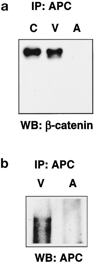
APC disappears in apoptotic HUVEC. (a) APC was immunoprecipitated from 250 μg lysate, and the immunoprecipitates from control (C), viable (V), and apoptotic (A) HUVEC were analyzed by Western blotting with the monoclonal antibody to β-catenin (571–781 C). Association of β-catenin and APC is abolished in apoptotic cells. (b) For analysis of the relative levels of APC under the different conditions, APC was immunoprecipitated from 750 μg of lysate from viable (V) and apoptotic (A) HUVEC with APC antibody (Ab-5), and the samples were separated on a 3% vertical agarose gel with Tris-borate-EDTA/0.1% SDS, transferred by inverse capillary transfer, and blotted with APC antibody Ab-1. APC is absent in apoptotic HUVEC.