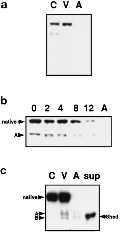
Figure 7.
VE-cadherin is shed from the cell surface during HUVEC apoptosis. (a) An antibody that recognizes the extracellular domain of VE-cadherin was used to evaluate control (C), viable (V), and apoptotic (A) cells. The extracellular epitope of VE-cadherin recognized by the antibody is absent in apoptotic cells. (b) After growth factor removal from HUVEC for 0, 2, 4, 8, and 12 h, pools of viable and apoptotic cells show a time-dependent loss of the extracellular epitope of VE-cadherin by Western analysis. A proteolytic fragment (A) is observed at 2 h and also decreases with time. (c) Analysis of supernatants (concentrated 10-fold) collected from apoptotic HUVEC cultures after 16 h without growth factors demonstrates that VE-cadherin is shed into the medium. Two fragments (A and B) are detected in viable and apoptotic cell lysates, and the approximately 90-kDa fragment (B) is also found in the supernatant.

