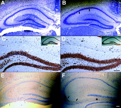
Figure 6.
Histological analysis of hippocampi of seizure positive B6.129-Kcnq2A306T/A306T knockin mice Nissl-stained coronal sections through the hippocampus of P42 aged B6.129-Kcnq2+/+ (A) and B6.129-Kcnq2A306T/A306T littermates (B) showing a lack of punctate staining in the CA1 pyramidal cell layer (arrows). Adjacent coronal Neu-N stained sections showing comparable hilar and stratum lacunosum interneurons (arrows) in B6.129-Kcnq2+/+ (C) and B6.129-Kcnq2A306T/A306T (D) mice. Insets illustrate the corresponding hippocampal section from which C and D were taken. Induction of NPY in hilar (*) and stratum lucidum (arrows) mossy fibres of Kcnq2A306T/A306T mice (F) following at least one generalized tonic–clonic seizure not seen in an age-matched control mouse (E). E and F are counterstained with haematoxylin. Scale bars, 100 μm.

