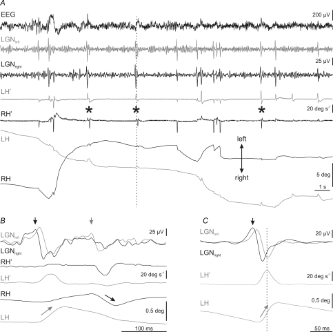
Figure 8. Temporal relationship between rapid eye movements and PGO waves during the transition period.
A, recording of electroencephalographic (EEG) activity, left (grey traces) and right (black traces) lateral geniculate nucleus (LGNleft, LGNright) activity, and horizontal eye velocity (LH′, RH′) and position (LH, RH) during a representative transition period. Rapid eye movements were associated to the occurrence of ponto-geniculo-occipital (PGO) waves at the LGN, and no PGO waves were recorded without the corresponding eye movement. Pairs of monocular abducting rapid eye movements of each eye are indicated by asterisks. B, expanded period at the time indicated by the vertical dotted line in A, showing a detail of LGN activities, and eye velocity and position during a pair of eye movements. Vertical arrows in LGN traces show the side (black, right; grey, left) on which primary PGO waves were recorded. Arrows on eye position indicate the eye that rotates in the abducting direction during primary PGO waves. C, average (n = 50) of left and right LGN activities during abducting movements of the left eye. The averaging was triggered by the peak velocity of the movement. Primary PGO wave, marked in this case by a black arrow, was always on the contralateral side with respect to the eye that performed the abducting movement.

