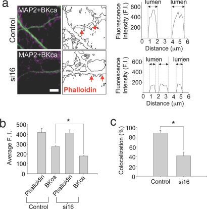
Fig. 3.
i16-specific siRNAs modify the differential distribution of BKCa channel protein in dendritic spines. (a Left) The merged confocal images of BKCa channel (magenta) and MAP2 (green) are shown. (Center) Outlines of AlexaFluor 488 phalloidin immunostaining are shown. Control and si16-treated cultures show a differential distribution of BKCa channel immunostaining. The line scan profile (Right) represents BKCa channel fluorescence intensity between where the red arrowheads point (Center). The higher BKCa channel staining was apparent in the lumen of control dendritic spines. The gray dotted lines in Right demarcate the edge of phalloidin staining which corresponds to the morphology of spines. (Scale bars: 5 μm.) (b) To quantify the differential distribution of BKCa channel in dendritic spines, we randomly selected dendritic spine heads based on phalloidin staining and measured the intensity of phalloidin and BKCa channel fluorescence intensities. There was no significant difference in phalloidin signal (control, 419.09 ± 43.01; si16, 415.20 ± 32.46). However, significant differences in BKCa channel staining were apparent (control, 274.47 ± 14.97; si16, 179.44 ± 4.74; n = 60; P < 0.001). (c) The correlation of dendritic spine and BKCa channel colocalization with si16 treatment. The incidence of colocalization observed for BKCa channels and phalloidin in dendritic spines of si16-treated neurons was dramatically reduced compared with control samples: 88.33% (41.67%, si16 treated culture; n = 60; P < 0.001). Data are mean ± SEM.

