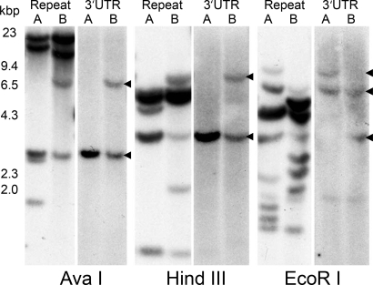
Fig. 3.
Southern blots of two diploid males (A and B) digested with three enzymes whose recognition sites are not in full-length bindin cDNA (AvaI, HindIII, and EcoRI). Shown is 10 μg of digested DNA per lane. Probes of the 3′ UTR show a maximum of two bands per individual, suggesting that oyster bindin is a single gene. Genomic DNA sequences indicate that each F-lectin repeat is bisected by an intron, and probes using a full-length repeat yield multiple bands. Of seven individuals analyzed, no two had identical patterns of hybridization with the repeat probe. HindIII-digested Lambda DNA size marker (kbp) is on the left.

