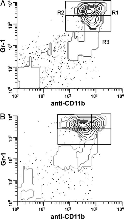
Fig. 3.
CS treatment of mice results in increases in all stages of granulocyte development. Bone marrow cells from mice implanted with cholesterol (A) or CS (B) were analyzed by flow cytometry by using the Gr-1/anti-CD11b/-Ly-6C system, with cells gated on Ly-6Cmed to eliminate contaminating monocytes. Regions corresponding to segmented neutrophils (R1), metamyelocytes and band cells (R2) and promyelocytes and myelocytes (R3) were analyzed. Representative data shown are from a control mouse (A) and a mouse with CS tablet implant (B) and are representative of typical myeloid changes in tablet implant experiments.

