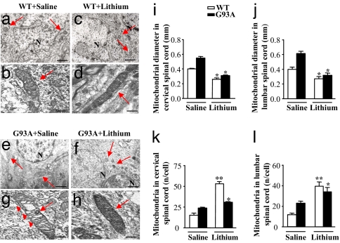
Fig. 3.
Effects of lithium administration on motor neurons mitochondria in vivo. (a–h) Representative pictures of mitochondria (arrows) in MN from the spinal cord of WT mice treated with saline (a and b) or lithium (c and d) and from G93A mice treated with saline (e and g) or lithium (f and h). (g) In G93A mice treated with saline, TEM shows mitochondrial vacuolization (arrowheads). (f and h) This vacuolization is consistently absent in mitochondria of G93A mice treated with lithium. (d and h–j) Lithium decreases the size of mitochondria both in WT and G93A mice (d and h, respectively) both in the cervical (i) and lumbar (j) spinal cord. (k and l) Lithium increases the number of mitochondria both in cervical (k) and lumbar (l) MN both in WT and G93A. Values are the mean ± SEM. Comparison between groups was made by using one-way ANOVA. *, P ≤ 0.05 compared with saline-treated mice. **, P ≤ 0.01 compared with saline treated mice. (Scale bars: a, c, e, and g, 1.8 μm; b, d, f, and h, 0.25 μm.)

