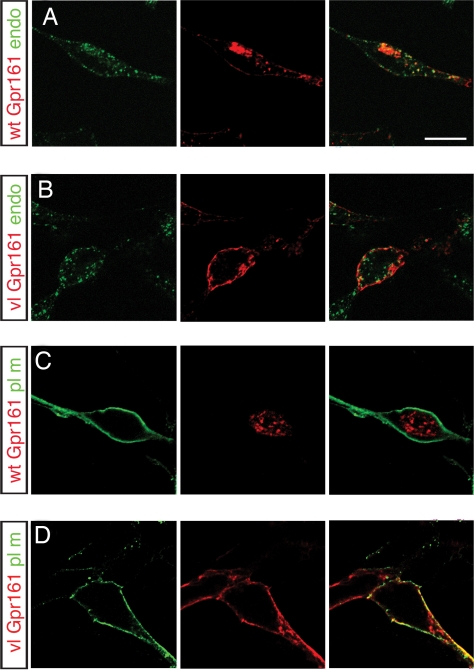
Fig. 3.
vl subcellular localization. Double labeling for WT (A and C) and vlGpr161 (B and D) with either the endosome marker, FITC-transferrin (A and B), or plasma membrane-targeted GFP (C and D) was performed for permeabilized transiently transfected HEK293T cells. wtGpr161 is localized to the endosome compartment, whereas vlGpr161 remains on the plasma membrane, consistent with the C-terminal tail truncation affecting receptor-mediated endocytosis of the Gpr161. Confocal microscopy of ≈0.5-μm optical sections through transfected cells is shown. (Scale bar: 10 μm.)

