Fig. 1.
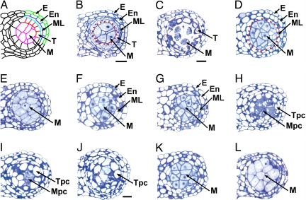
Ectopic expression of TPD1 causes abnormal anther cell differentiation, and TPD1 signaling requires EMS1. Semithin sections show one lobe from anthers. (Scale bars: 20 μm.) B, D, F–H, and K; C, E, I, and L have the same magnification. (A) Diagram of a mature wild-type anther shows epidermis (E), endothecium (En), middle layer (ML), tapetum (T), and microsporocytes (M). (B) A wild-type anther at stage 5. Enclosed by red dotted line are microsporocytes. (C) A wild-type anther at stage 6 shows strongly stained tapetal layer and isolated microsporocytes. (D) An ems1 anther at stage 5 lacks the tapetum layer but has excess microsporocytes (enclosed by red dotted line). (E) An ems1 anther at stage 6 lacks the tapetum layer. Microsporocytes are abnormally enlarged and not isolated. (F) A tpd1 anther at stage 5 has the same phenotype as that of ems1 (D). (G) An ems1 tpd1 double-mutant anther at stage 5 shows the same phenotypes as those of ems1 (D) and tpd1 (F). (H–J) Anthers of CaMV35S::TPD1 transgenic plants. (H) A stage-5 anther shows vacuolated cells in place of a normal tapetum (tapetum-positioned cells, Tpc) and degenerating cells instead of normal microsporocytes (microsporocyte-positioned cells, Mpc). (I) A stage-6 anther exhibits completely vacuolated Tpc and degenerating Mpc. (J) An anther after stage 6 shows the mix of completely vacuolated Tpc and Mpc. (K and L) Anthers from CaMV35S::TPD1 ems1 plants at stage 5 (K) and 6 (L) exhibit identical phenotypes to ems1 (D and E).

