Fig. 3.
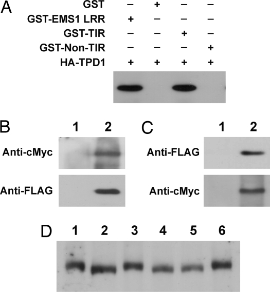
In vitro and in vivo interaction between TPD1 and EMS1 and TPD1 induces the phosphorylation of EMS1. (A) TPD1 interacts with EMS1 in GST pull-down assay. Three micrograms of GST and GST fusion proteins were used to pull down the same amount of crude protein (200 μg) extracts containing HA-TPD1, respectively. (B and C) TPD1 interacts with EMS1 in planta in coimmunoprecipitation assay. (B) EMS1-cMyc (Upper) and TPD1-FLAG (Lower) were detected in EMS1::EMS1-cMyc TPD1::TPD1-FLAG double-transgenic plants, respectively, by Western blot. (1) Wild type. (2) EMS1::EMS1-cMyc TPD1::TPD1-FLAG double-transgenic plant. (C) TPD1-FLAG was detected when proteins from EMS1::EMS1-cMyc TPD1::TPD1-FLAG double-transgenic plants were immunoprecipitated with an anti-cMyc antibody (Upper). EMS1-cMyc was also detected when the same proteins were immunoprecipitated with an anti-FLAG antibody (Lower). (1) TPD1::TPD1-FLAG (Upper) and EMS1::EMS1-cMyc (Lower) single-transgenic plants. (2) EMS1::EMS1-cMyc TPD1::TPD1-FLAG double-transgenic plant. (D) TPD1 binding induces EMS1 phosphorylation. Lanes 1, 3, 4, and 6: EMS1::EMS1-cMyc transgenic plants; lanes 2 and 5: EMS1::EMS1-cMyc tpd1 transgenic plants, in which TPD1 is not present. Proteins in lanes 4 and 5 were treated with calf intestinal alkaline phosphatase (CIP). EMS1-cMyc protein from EMS1::EMS1-cMyc tpd1 plants (lane 2) migrated faster than those from EMS1::EMS1-cMyc plants (lanes 1 and 3). A shift in mobility of EMS1-cMyc did not occur (lane 4) after the CIP treatment (lanes 5 and 6 are controls).

