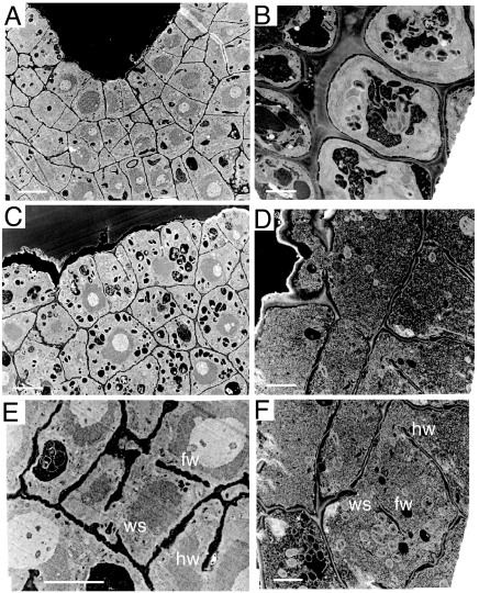
Fig. 2.
Electron micrographs showing defective cell walls in the rsh/rsh mutant. (A, C, and E) Images of heart-stage embryo sections. (B, D, and F) Images of 3-day-old root transverse sections. The mutant (C–F) compared with the wild type (A and B) has incomplete cell walls. fw, floating walls; hw, hanging walls; ws, wall stubs. [Scale bars: 10 μm (A, C, and E) and 4 μm (B, D, and F).]

