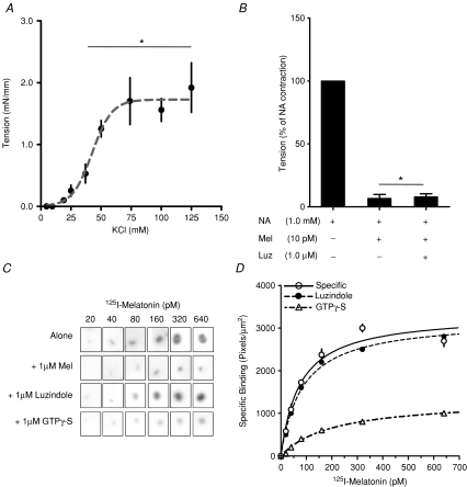
Figure 1. Detection of a functional melatonin receptor in sheep fetal cerebral arteries at 90% of gestation.
A, contractile response of cerebral arteries to potassium chloride (KCl). Values are means ±s.e.m. of tension. *Different to basal tension (P < 0.05; ANOVA and Newman–Keuls). B, effect of melatonin (Mel) on maximal contraction induced by 1 mm noradrenaline (NA) in presence or absence of luzindole (Luz). Values are percentage of maximal NA contraction. *Different to NA contraction (P < 0.05; ANOVA and Newman–Keuls). C, specific 2-[125I]iodomelatonin saturation binding by whole-mount autoradiography of cerebral artery rings sections exposed to increasing concentrations of 2-[125I]iodomelatonin. Each incubation condition (2-[125I]iodomelatonin alone, plus 1 μm cold melatonin; plus 1 μm luzindole and 1 μm GTPγ-S), is indicated in the figure. D, saturation isotherms of specific 2-[125I]iodomelatonin binding in cerebral arteries in presence of luzindole or GTPγ-S.

