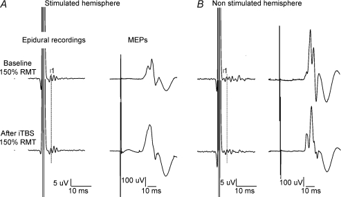
Figure 3. Corticospinal volleys and motor evoked potentials evoked by single pulse magnetic stimulation in baseline conditions and after right motor cortex intermittent theta burst stimulation (iTBS) in subjects 3 after stimulation of the ipsilateral (left panel) and contralateral (right panel) hemisphere.
Each trace is the average of 20 sweeps. A, magnetic stimulation evokes three descending waves. The latency of the earliest (I1) wave is indicated by the vertical line. After iTBS, the size of the I2 and I3 waves is increased (F1,18 = 9.04, P = 0.008), the amplitude of the I1 wave is unchanged (F1,18 = 0.137, P = 0.716). The amplitude of MEP is significantly increased after iTBS (F1,18 = 6.35, P = 0.021). B, magnetic stimulation evokes several descending waves. After iTBS, the size of the later waves is significantly decreased (F1,18 = 10.74, P = 0.004), and the amplitude of the I1 wave is unchanged (F1,18 = 1.3, P = 0.268). The amplitude of MEP is decreased after iTBS, but the change is not significant (F1,18 = 0.59, P = 0.453).

