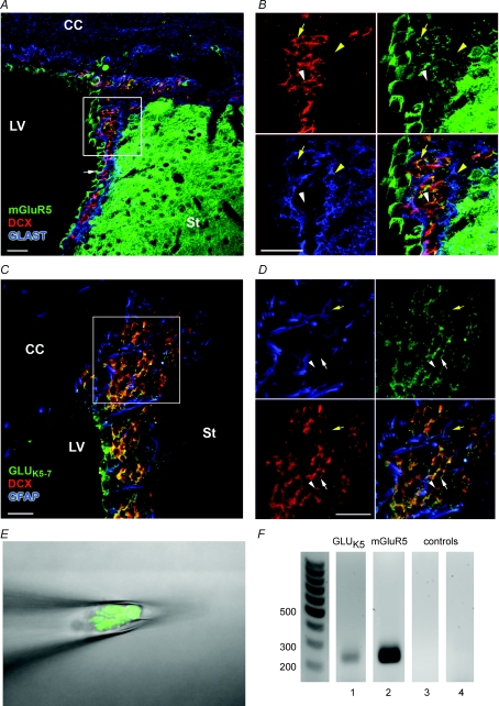
Figure 1. SVZ neuroblasts express mGluR5 and GLUK5–7-containing kainate receptors.
A and B, photographs of immunostaining for mGluR5 (green), DCX (red) and GLAST (blue) in a coronal section from a P30 mouse brain at low (A) and high magnification (B). mGluR5 (green) is expressed by neuroblasts (red, yellow arrow) and ependymal cells (white arrow in A), but not by GLAST-positive cells, i.e. astrocytes (blue, yellow arrowhead). C and D, photographs of immunostaining for GLUK5–7-containing receptors (green), DCX (red) and GFAP (blue) in a coronal section from a P30 mouse brain at low (C) and high magnification (D). GLUK5–7-containing receptors are expressed by neuroblasts (red, white arrow) and in a few GFAP cells (blue, yellow arrow). Some neuroblasts do not stain for GLUK5–7-containing receptors (white arrowhead). Scale bars, 20 μm. E and F, agarose gel electrophoresis (F) demonstrating RT-PCR amplification of GLUK5 (lane ‘1’) and mGluR5 (lane ‘2’) mRNA isolated during pipette aspiration of 10 GFP-fluorescent cells, i.e. neuroblasts, shown in E. GLUK5 and mGluR5 were not detected in bath solution (lane ‘3’ for GLUK5 and lane ‘4’ for mGluR5) controls.

