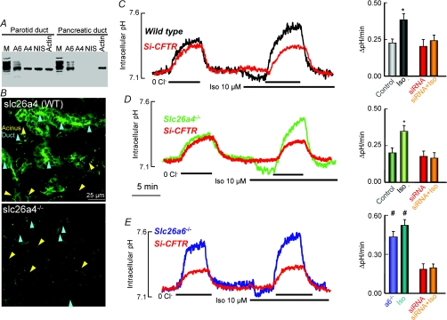
Figure 5. Slc26a6, but not Slc26a4, mediates luminal Cl−/HCO3− exchange in the parotid duct.
A shows RT-PCR analysis of Slc26a6 (A6), Slc26a4 (A4), and Na+–I− symporter (NIS) in parotid and pancreatic ducts. Pancreatic duct is used as a control for Slc26a4 and NIS and actin is loading control. M stands for markers. B shows immunolocalization of Slc26a4 in submandibular section. The lower image is with a submandibular gland from Slc26a4−/− mice. Acini and ducts are marked by yellow and cyan arrowheads, respectively. Note that Slc26a4 is expressed in the luminal membrane of the duct and is not expressed in the acini. In C–E, Cl−/HCO3− exchange activity was measured in sealed ducts by equilibrating the ducts in HCO3−-buffered media for at least 15 min before alternately incubating them in Cl−-containing and Cl−-free medium before and after stimulation with 10 μm isoproterenol (Iso). The ducts were cultured for 36–48 h and treated with scrambled (control) or CFTR siRNA (red traces). Ducts were microdissected from wild-type (C), Slc26a4 (D) or Slc26a6 (E) deficient mice. The columns are the mean ±s.e.m. of 6–8 ducts from 3 or 4 mice of each line. Note that deletion of Slc26a4 has no effect on basal or stimulated Cl−/HCO3− exchange activity, while deletion of Slc26a6 enhances basal activity that is not further stimulated by Iso. KD of CFTR inhibited only the stimulated Cl−/HCO3− exchange activity in wild-type and Slc26a4−/− ducts, whereas KD of CFTR inhibited the enhanced basal Cl−/HCO3− exchange activity in Slc26a6−/− ducts.

