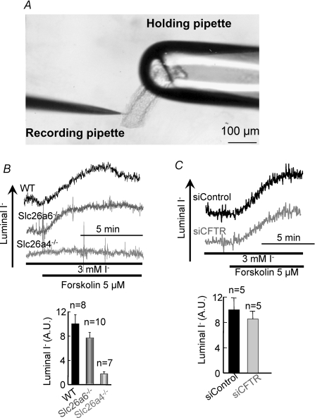
Figure 6. Slc26a4, but not Slc26a6, mediates luminal I− secretion in the parotid duct.
A shows an image of sealed ducts held by a large-bore holding pipette and penetrated with a recording pipette. In B, wild-type (dark trace and columns), Slc26a6−/− (grey trace and columns), and Slc26a4−/− (light grey trace and columns) sealed parotid ducts were incubated with 3 mm I− for 15 min before stimulation of I− secretion with 5 μm forskolin. The columns are the mean ±s.e.m. of the indicated number of experiments. Note that activation of I− secretion is observed with wild-type and Slc26a6−/− ducts, but not with Slc26a4−/− ducts. In C, wild-type ducts treated with scrambled (dark trace and columns) or CFTR siRNA (grey trace and columns) were used to measure stimulated I− secretion. The columns show the mean ±s.e.m. of 5 experiments. AU, arbitrary units. Note that KD of CFTR that reduced Cl−/HCO3− exchange activity in these ducts (Fig. 5C) had no effect on I− secretion.

