Figure 1.
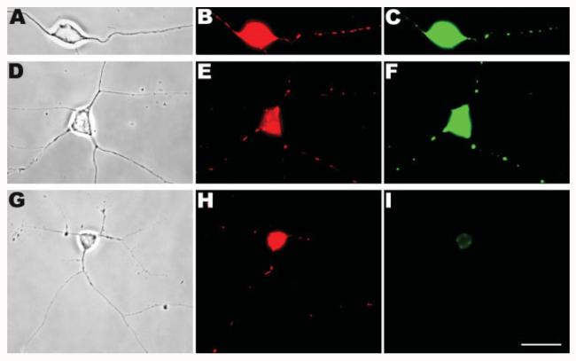
In situ detection of BrdU-incorporated mtDNA. Chick peripheral neurons were incubated with BrdU and the incorporation into mtDNA was detected by anti-BrdU immunocytochemistry. Phase contrast images (A,D,G), epifluorescence images of MitoTracker-labeled mitochondria (B,E,H), and of BrdU-labeled DNA (C,F,I) are shown here. After 15–20 h of labeling, BrdU signals were readily detected in mitochondria along the length of the axons (A–C). A similar pattern of BrdU localization was obtained even when the labeling period was decreased to 3 h (D–F). However, preincubation with ddC (G–I) resulted in no BrdU signals in the axons even after 15–20 h of BrdU staining and only a low level of background in the cell body (I). Note that due to the amplification with the double-precipitation procedure, the BrdU signals appeared to almost completely overlap with Mitotracker signals rather than being discrete foci within mitochondria. In addition, because of the immediate proximity to the cell body and long duration of BrdU exposure in (A–C), most of the mitochondria seen are BrdU-positive. Scale bar, 10 μm.

