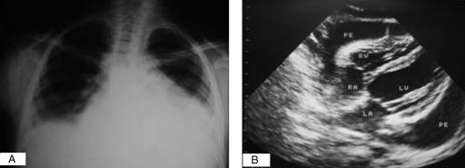
Figure 1).
A Chest x-ray of a patient with a large pericardial effusion showing gross cardiomegaly. B Two-dimensional echocardiography showing a large pericardial effusion and collapse on the free wall of the right atrium. LA Left aorta; LV Left ventricle; RA Right aorta; RV Right ventricle; PE Pericardial effusion

