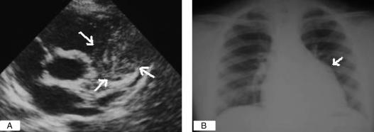
Figure 2).
Hydatid cyst mimicking a solid mass. A Echocardiographic image of the parasternal short axis showing a round lesion (arrows) with mixed echogenic circles and serpentine structures within the matrix, representing collapsed membranes on the right ventricle outlet tract and treaded main pulmonary artery inside the pericardial cavity. B Chest x-ray showing a bulge (arrow) along the left cardiac border where the mass was located

