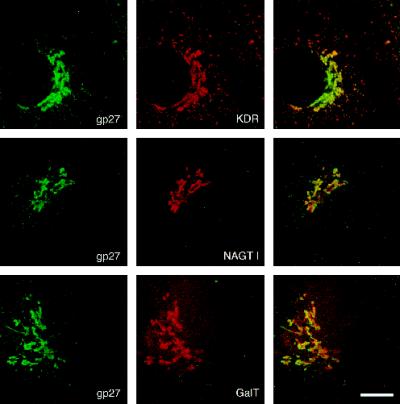
Figure 1.
Comparison between gp27 and Golgi marker proteins. Double immunofluorescence stainings of Vero cells were analyzed by confocal laser scanning microscopy. Marker proteins were chosen to represent subcompartments of the Golgi apparatus: KDEL receptor for CGN and cis-Golgi, NAGT I for medial-Golgi, and GalT (GTL2 antibody) for trans-Golgi and TGN. Note that the localization of KDEL receptor and gp27 is very similar, although intensities are different. NAGT I and, even more, GalT show Golgi patterns shifted against gp27. Bar, 10 μm.

