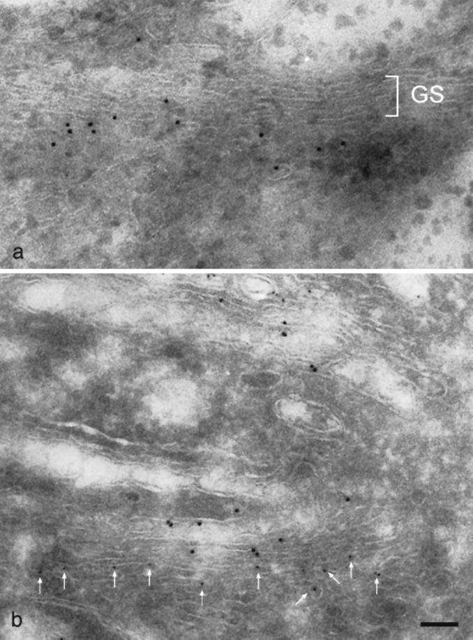
Figure 2.
Ultrastructural localization of gp27 to the cis side of the Golgi apparatus. (a) Single labeling of HeLa cells with gp27 followed by protein A-colloidal gold (10 nm). Golgi stacks (GS) usually comprise three cisternae, and labeling for gp27 is highly polarized. (b) Comparison between the trans-Golgi marker protein GalT (10 nm, rabbit antiserum N10) and gp27 (5 nm). Segregation between these two proteins identifies the gp27-containing cisternae as cis-Golgi. Bar, 100 nm.

