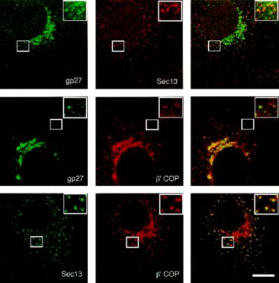
Figure 3.
Localization of gp27 to coated transport structures between ER and Golgi. Double immunofluorescence stainings of Vero cells were analyzed by confocal laser-scanning microscopy. Sec13/COPII and β′-COP/COPI show some coincident staining with gp27, which is more evident in the insets. For comparison, the much more obvious closely associated staining between COPII and COPI is shown in the lower panel. Bar, 10 μm.

