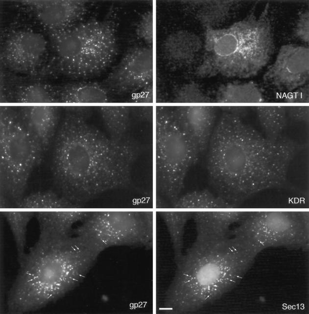
Figure 5.
Subcellular distribution of gp27 after brefeldin A treatment. Resident glycosylation enzymes such as NAGT I stain nuclear envelope and reticular cytoplasmic ER structures, but gp27 distribution is different. A striking coincidence of staining is observed with KDEL receptor, and many of the brefeldin A–induced punctated structures are coincident with Sec13/COPII. Bar, 10 μm.

