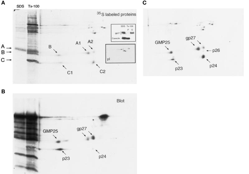
Figure 7.
Coimmunoprecipitation of p24 family proteins with gp27 affinity-purified antibodies. [35S]Methionine was used for labeling HeLa cells overnight, and lysis was routinely done with the nonionic detergent TX-100. Under these conditions, gp27 was efficiently solubilized (a, inset; p, pellet; S, supernatant), whereas large detergent-resistant structures containing caveolin, a protein residing mostly in the CGN and the cis-Golgi as well as on plasma membrane (Dupree et al., 1993; Parton, personal communication), were sedimented and would not be present in the starting lysate for immunoprecipitation. Immunoprecipitates obtained with gp27 antibodies were analyzed by 1D SDS-PAGE (a, lanes SDS and TX-100) and isoelectric focusing followed by SDS-PAGE (a–c). Under denaturing conditions (a, SDS), one prominent band at 30 kDa corresponding to gp27 is resolved. Using TX-100 (b, Tx-100), three major bands in the 20- to 30-kDa range were observed (A–C). 2D gel analysis resolved this pattern into five different spots: A1, A2, B, C1, and C2 (a). The same 2D gel loaded with 35S-labeled immunoprecipitate and unlabeled Golgi-enriched subcellular fraction is shown in a and b. After detection of radioactively labeled proteins (a), the membrane was probed with antibodies against p24 family proteins (b). Alignment of the two patterns identified coprecipitated proteins as different p24 family members. Protein A-peroxidase was applied for detection of primary antibodies against p24 family proteins, which also bound to IgGs of the gp27 antibodies used for immunoprecipitation, giving rise to the additional strong staining patterns apparent in b. Some additional spots of higher molecular mass are also apparent and are likely to be unspecific contaminants, because they are also seen when using preimmune serum (pI) or rabbit IgG (a, inset). 2D gels loaded with labeled immunoprecipitate without added Golgi fraction were of sufficient resolution to enable quantification (c).

