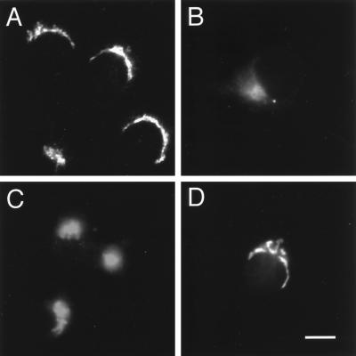
Figure 2.
Perinuclear labeling for VAMP4 is detected in cell lines and primary cultures. Cells were fixed with 4% paraformaldehyde, permeabilized with saponin, and stained using affinity-purified anti-VAMP4 polyclonal antibodies and Texas Red–conjugated anti-rabbit IgG secondary antibody before processing for indirect immunofluorescence microscopy. (A) NRK; (B) COS-7; (C) NGF-differentiated PC12; (D) 12 d in vitro embryonic hippocampal cultures. Note that there is no VAMP4 staining observed in the processes of differentiated PC12 cells or embryonic hippocampal cultures. Bar: A–C, 20 μm; D, 25 μm.

