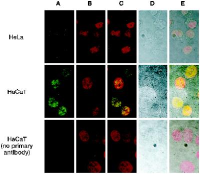
Figure 5.
hRAD9 is located in the nucleus. Confocal immunofluorescence and light microscopy was performed on <50% confluent HeLa cells and 90% confluent HaCaT cells. Cells were fixed and probed with α-hRAD9 chicken polyclonal antibodies, followed by a fluorescently labeled anti-chicken IgY secondary antibody (A). DNA was visualized by staining with propidium iodide (B). Images from A and B were superimposed (C). The cellular borders of the HeLa and confluent HaCaT cells were visualized by light microscopy (D), and the light and fluorescent images were superimposed (E).

