Figure 1.
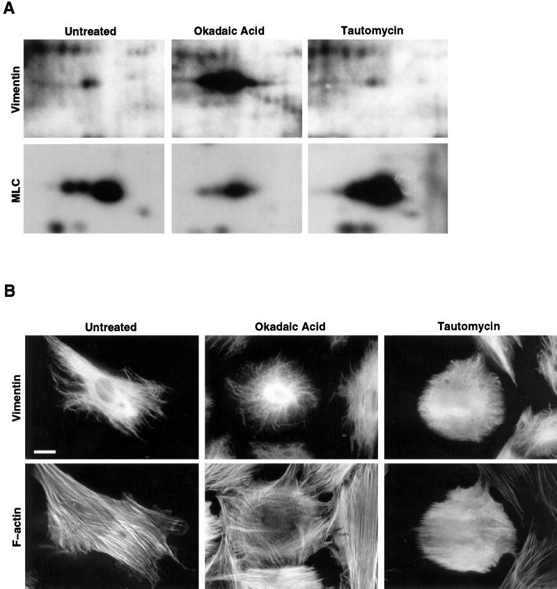
Analysis of phosphoproteins and the cytoskeleton of OA- and TAU-treated cells. (A) Equal numbers of Hs68 fibroblasts, grown on 12-mm glass coverslips, were treated with OA or TAU and metabolically labeled with [32P]H3PO4, and the totality of cellular proteins was separated by two-dimensional gel electrophoresis as described in MATERIALS AND METHODS. Shown are portions of the autoradiograms containing vimentin (upper panels) and myosin light chain (lower panels). Untreated, phosphoproteins of untreated cells labeled for 60 min; Okadaic Acid, phosphoproteins of cells treated with 1 μM OA during the 60-min labeling period; Tautomycin, phosphoproteins of cells treated with 10 μM TAU during 2 h and the subsequent 60-min labeling period. Autoradiographs were exposed at −80°C for 2 h for vimentin and for 6 h for MLC. (B) Hs68 fibroblasts were either left untreated or incubated with OA (1 μM for 45 min) or TAU (10 μM for 2 h). Subsequently they were fixed in formalin and costained for vimentin and F-actin using mAb V9 and Bodipy-phalloidin, respectively. Bar, 5 μm.

