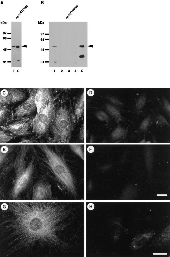
Figure 2.
Subcellular distribution of the B55 subunit in Hs68 fibroblasts. (A) Approximately 20 μg of total cell extracts from Hs68 fibroblasts were electrophoretically separated (lane T) and immunoblotted; ∼10 ng of recombinant B55α (lane C) were included as positive control. Immunoblots were decorated with affinity-purified Ab55473/448. (B) Hs68 fibroblasts were differentially extracted for soluble cytoplasm (lane 1), insoluble cytoplasm (lane 2), nucleoplasm (lane 3), and the nuclear pellet also containing microfilament and IFs (lane 4). Twenty micrograms of the soluble (lanes 1 and 3) and equal volumes of the insoluble (lanes 2 and 4) fractions were electrophoretically separated and immunoblotted using Ab55recomb. An identical subcellular distribution was obtained using affinity-purified Ab55473/448. Note that both antibodies recognize different proteolytic fragments of recombinant B55 (lane C), namely an amino terminus with Ab55recomb and a carboxyl terminus for Ab55473/448. (C–H) Hs68 fibroblasts were grown on glass coverslips and formalin fixed. Cells were stained for B55 using either Ab55473/448 (C and D) or Ab55recomb (E–H). In D, F, and H, antibodies were preincubated with recombinant B55 as described in MATERIALS AND METHODS. The images resulting from the same antibody staining were acquired and treated identically. (G and H) Confocal section of ∼180 nm through the middle of the nucleus. Bar, 5 μm.

