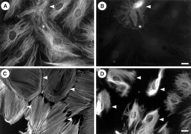
Figure 5.
Microtubule and microfilament networks are unaffected in B55-depleted cells. Hs68 fibroblasts were microinjected with pECE-B55as and cultured for 12 h as in Figure 4. (A and B) Cells were then stained for tubulin (A). Arrowheads indicate microinjected cell as identified by costaining against the marker antibody (B). (C and D) Costaining for F-actin (C) and vimentin (D), respectively. Here the corresponding guinea pig marker antibody (revealed with amino-methylcoumarin) is not shown but indicated by arrowheads. In each case ∼50 cells were microinjected, and no effect on microtubule or actin distribution was observed. Bars, 5 μm.

