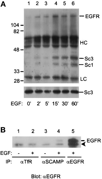
Figure 6.
Time course of SCAMP3 phosphorylation. (A) NeoR cells were stimulated with 100 ng/ml EGF for 0, 2, 5, 15, 30, or 60 min, lysed in CHAPS buffer, and immunoprecipitated with anti-SCAMP (7C12). Samples were Western blotted for phosphotyrosine (αP-Tyr, upper panel) or for SCAMP3 (3γ, lower panel). (B) EGF-inducible association of SCAMPs with EGFR. NeoR cells were either unstimulated (lanes 1 and 3) or EGF stimulated (lanes 2, 4, and 5), and then immunoprecipitated with a control antibody (anti-TfR, lanes 1 and 2), anti-SCAMP (7C12, lanes 3 and 4), or anti-EGFR (lane 5). Samples were resolved by 7.5% SDS-PAGE, transferred to nitrocellulose, Western blotted for EGFR, and visualized by ECL. The arrowhead indicates the position of a nonspecific band of ∼150 kDa.

